Exploring Digital Ultrasound Machines: Key Features & Tech
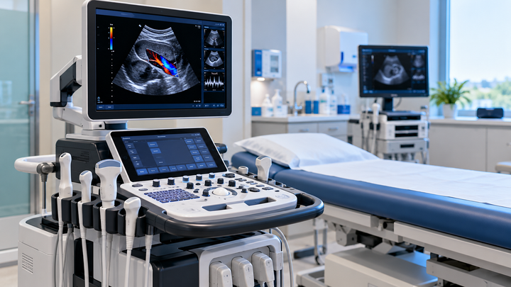
Digital ultrasound machines are medical imaging systems that use high-frequency sound waves and digital signal processing to create real-time images of internal organs, tissues, blood vessels, and developing fetuses.
These systems are widely used because they provide non-invasive imaging without ionizing radiation and support rapid clinical assessment.
Modern digital ultrasound technology combines advanced transducers, image processing algorithms, software-based measurements, and network connectivity to improve image quality, workflow efficiency, and diagnostic capabilities across many medical specialties.
Quick Facts About Digital Ultrasound Machines
| Feature | Details |
|---|---|
| Technology | High-frequency sound wave imaging |
| Imaging Type | Real-time diagnostic imaging |
| Radiation Use | No ionizing radiation |
| Common Applications | Obstetrics, cardiology, radiology, emergency medicine |
| Main Components | Transducer, processor, display, software |
| Image Modes | B-mode, Doppler, M-mode, 3D and 4D |
| Portability | Available in cart-based and portable formats |
| Typical Users | Sonographers, physicians, radiologists |
What Are Digital Ultrasound Machines?
Digital ultrasound machines are diagnostic imaging devices that transmit sound waves into the body and receive returning echoes. These echoes are processed digitally to generate images of internal anatomical structures.
Unlike older analog systems, digital ultrasound platforms use advanced computing technologies for signal processing, image enhancement, measurements, storage, and workflow integration. This allows clinicians to visualize soft tissues, organs, blood flow, and fetal development with improved clarity and efficiency.
Why Digital Ultrasound Technology Matters
Ultrasound remains one of the most widely used imaging technologies because it offers real-time visualization without exposing patients to ionizing radiation.
Key reasons digital ultrasound systems are important include:
- Real-time imaging capabilities
- Non-invasive diagnostic evaluation
- Wide range of clinical applications
- Improved workflow through digital processing
- Enhanced image storage and sharing
- Portable deployment options
- Support for bedside assessments
- Compatibility with healthcare information systems
Digital platforms have expanded ultrasound applications from traditional radiology departments to emergency rooms, operating theaters, outpatient clinics, and mobile healthcare environments.
Understanding the Digital Ultrasound Concept
Simple Explanation
A digital ultrasound machine works similarly to sonar technology. The transducer sends sound waves into the body. When these waves encounter organs, tissues, or blood vessels, they bounce back as echoes.
The system analyzes these returning echoes using digital processors and converts them into detailed visual images displayed on a monitor. Different tissue types reflect sound differently, allowing clinicians to distinguish anatomical structures.
How Digital Ultrasound Machines Work
Sound Wave Generation
The transducer contains piezoelectric elements that generate high-frequency sound waves when electrical energy is applied.
Sound Wave Transmission
The sound waves travel through tissues and body fluids. Ultrasound gel helps improve transmission by reducing air gaps between the transducer and skin.
Echo Reception
Reflected sound waves return to the transducer after interacting with tissues and anatomical structures.
Digital Signal Processing
The machine converts received echoes into digital signals and applies image enhancement algorithms.
Image Formation
Software reconstructs the processed signals into real-time diagnostic images.
Clinical Interpretation
Healthcare professionals analyze the images to support diagnosis, monitoring, and treatment planning.
Digital Ultrasound Workflow Table
| Workflow Stage | Function |
|---|---|
| Patient Preparation | Position patient and select exam type |
| Probe Selection | Choose suitable transducer |
| Signal Transmission | Send sound waves into tissues |
| Echo Detection | Capture returning signals |
| Digital Processing | Convert echoes into images |
| Image Optimization | Adjust gain and parameters |
| Analysis | Perform measurements and assessment |
| Reporting | Document findings and store images |
Digital Ultrasound Machine Architecture
Transducer Assembly
The transducer serves as both the transmitter and receiver of ultrasound signals. Different probe designs are used for specific anatomical applications.
Beamforming System
Beamforming electronics control how ultrasound waves are transmitted and received, improving image resolution and focus.
Digital Signal Processor
This component processes incoming echo data and performs image reconstruction calculations.
User Interface
The operator console includes controls, keyboards, touchscreens, and imaging adjustments.
Display System
High-resolution monitors provide real-time visualization of anatomical structures.
Data Storage Module
Images, measurements, and reports can be stored digitally for review and documentation.
Connectivity System
Modern systems support integration with hospital networks and imaging archives.
Main Components of Digital Ultrasound Machines
| Component | Function |
|---|---|
| Transducer | Sends and receives sound waves |
| Beamformer | Controls ultrasound beam direction |
| Signal Processor | Converts echoes into image data |
| Display Monitor | Shows diagnostic images |
| Control Panel | Allows user adjustments |
| Storage System | Saves images and reports |
| Software Platform | Provides imaging tools |
| Network Interface | Enables data sharing |
Types of Digital Ultrasound Machines
Cart-Based Ultrasound Systems
Large systems commonly used in hospitals and imaging centers.
Advantages:
- Advanced imaging capabilities
- Multiple probe compatibility
- Expanded clinical applications
Portable Ultrasound Systems
Compact units designed for mobility within healthcare facilities.
Advantages:
- Easy transport
- Faster bedside assessment
- Flexible deployment
Handheld Ultrasound Devices
Highly compact devices suitable for point-of-care imaging.
Advantages:
- Lightweight design
- Rapid accessibility
- Immediate clinical evaluation
Specialized Ultrasound Systems
Designed for dedicated applications such as cardiology or obstetrics.
Advantages:
- Optimized workflows
- Specialized software packages
- Application-focused imaging tools
Digital Ultrasound Machine Type Comparison
| Type | Mobility | Typical Use | Main Strength |
|---|---|---|---|
| Cart-Based | Low | Hospitals | Advanced imaging |
| Portable | Medium | Clinics and wards | Flexibility |
| Handheld | High | Point-of-care imaging | Accessibility |
| Specialized | Varies | Dedicated departments | Clinical optimization |
Requirements for Effective Digital Ultrasound Imaging
Successful ultrasound imaging depends on multiple technical and operational factors.
Key requirements include:
- Appropriate transducer selection
- Proper patient positioning
- Adequate acoustic coupling gel
- Skilled image acquisition
- Regular equipment calibration
- Reliable power supply
- Software updates and maintenance
- Compliance with clinical protocols
Digital Ultrasound Machines vs Traditional Imaging Technologies
| Feature | Digital Ultrasound | CT Scan | MRI |
|---|---|---|---|
| Radiation Exposure | None | Uses ionizing radiation | None |
| Real-Time Imaging | Yes | Limited | Limited |
| Portability | Available | Typically fixed | Fixed installation |
| Soft Tissue Imaging | Good | Good | Excellent |
| Bedside Use | Possible | Rare | Not practical |
| Blood Flow Assessment | Doppler capable | Limited | Available in specific protocols |
Digital ultrasound is often selected when real-time imaging, portability, and radiation-free assessment are priorities.
Benefits and Advantages of Digital Ultrasound Machines
- Real-time image visualization
- No ionizing radiation exposure
- Broad clinical versatility
- Faster diagnostic workflow
- Digital image storage capabilities
- Portable deployment options
- Enhanced image processing
- Doppler blood flow assessment
- Improved measurement accuracy
- Integration with healthcare systems
These advantages contribute to widespread use across diagnostic, monitoring, and procedural applications.
Industry Applications of Digital Ultrasound Machines
Obstetrics and Gynecology
Ultrasound is commonly used to monitor fetal development, evaluate pregnancy progression, and assess reproductive health.
Cardiology
Cardiac ultrasound supports evaluation of heart structures, valve function, and blood flow dynamics.
Radiology
Radiologists use ultrasound to examine abdominal organs, soft tissues, and vascular structures.
Emergency Medicine
Point-of-care ultrasound assists rapid clinical assessment in emergency settings.
Musculoskeletal Imaging
Clinicians evaluate tendons, muscles, ligaments, and joint structures using ultrasound imaging.
Vascular Medicine
Doppler ultrasound helps assess blood flow and vascular conditions.
Interventional Procedures
Ultrasound guidance supports various minimally invasive procedures and needle placement.
Digital Ultrasound Applications Table
| Medical Field | Common Application |
|---|---|
| Obstetrics | Pregnancy monitoring |
| Cardiology | Echocardiography |
| Radiology | Abdominal imaging |
| Emergency Medicine | Rapid assessment |
| Vascular Medicine | Blood flow evaluation |
| Musculoskeletal Care | Tendon and joint imaging |
| Interventional Medicine | Procedure guidance |
Industry Standards and Safety
Digital ultrasound equipment is developed and used within recognized medical device and healthcare standards frameworks.
Important areas include:
- IEC 60601 medical electrical equipment safety requirements
- IEC 62304 medical device software lifecycle processes
- DICOM compatibility for medical image management
- Quality management practices based on ISO 13485
- Clinical safety guidelines for ultrasound exposure
Healthcare providers should ensure ultrasound examinations are performed by qualified professionals using established clinical protocols.
Common Challenges and Solutions
| Challenge | Practical Solution |
|---|---|
| Operator dependency | Ongoing training and education |
| Image artifacts | Proper scanning techniques |
| Limited penetration in some patients | Appropriate probe selection |
| Data management complexity | Integrated digital workflows |
| Equipment maintenance needs | Scheduled preventive maintenance |
| Workflow variability | Standardized imaging protocols |
How to Choose a Digital Ultrasound Machine
| Selection Factor | Evaluation Criteria |
|---|---|
| Clinical Specialty | Cardiology, obstetrics, radiology, or general imaging |
| Image Quality | Resolution and visualization requirements |
| Portability Needs | Fixed, portable, or handheld deployment |
| Probe Compatibility | Supported transducer types |
| Software Features | Measurements and reporting tools |
| Connectivity | Network and archive integration |
| Workflow Requirements | Clinical environment needs |
| Future Expansion | Upgrade and scalability options |
Maintenance Best Practices
Regular maintenance helps maintain imaging performance and operational reliability.
Recommended practices include:
- Routine transducer inspections
- Cleaning according to manufacturer instructions
- Software maintenance and updates
- Display quality checks
- Data backup procedures
- Electrical safety inspections
- Scheduled calibration verification
- Preventive maintenance programs
Clinical users should follow manufacturer guidance and institutional maintenance protocols.
Limitations of Digital Ultrasound Machines
Although ultrasound offers many advantages, it has limitations.
Image quality can be affected by operator experience, patient anatomy, and scanning conditions. Ultrasound generally has limited ability to image structures obscured by bone or air-filled organs. Some complex clinical situations may require complementary imaging methods such as CT or MRI for a more comprehensive evaluation.
Additionally, image interpretation requires appropriate clinical training and expertise.
Future Trends in Digital Ultrasound Technology
Several developments continue to shape modern ultrasound systems.
Emerging trends include:
- Artificial intelligence-assisted image analysis
- Improved portable imaging platforms
- Advanced 3D and 4D visualization
- Enhanced automation tools
- Cloud-connected workflow systems
- Improved transducer technology
- Faster image processing hardware
- Expanded point-of-care ultrasound adoption
These innovations aim to improve workflow efficiency, image quality, and accessibility while supporting clinical decision-making.
Frequently Asked Questions About Digital Ultrasound Machines
What is a digital ultrasound machine?
A digital ultrasound machine is a medical imaging system that uses sound waves and digital processing to create real-time images of internal body structures. It is commonly used for diagnosis, monitoring, and procedural guidance.
Do digital ultrasound machines use radiation?
No. Ultrasound imaging uses high-frequency sound waves rather than ionizing radiation, making it different from imaging methods such as CT scans.
What is the role of a transducer?
The transducer generates ultrasound waves and receives returning echoes. It is one of the most important components of an ultrasound system.
What is Doppler ultrasound?
Doppler ultrasound measures and visualizes blood flow within vessels and the cardiovascular system. It is widely used in vascular and cardiac imaging.
Are portable ultrasound machines reliable?
Modern portable systems can provide high-quality imaging for many clinical applications. Their suitability depends on the intended use and required imaging capabilities.
What medical specialties use ultrasound most often?
Common specialties include obstetrics, cardiology, radiology, emergency medicine, vascular medicine, and musculoskeletal care.
Why is ultrasound gel necessary?
Ultrasound gel improves sound wave transmission between the transducer and the skin by eliminating air gaps that can interfere with imaging.
Can ultrasound guide medical procedures?
Yes. Ultrasound is frequently used to guide needle placement and other minimally invasive procedures by providing real-time visualization.
What is the difference between 3D and 4D ultrasound?
3D ultrasound produces three-dimensional images, while 4D ultrasound adds real-time movement to those images.
How often should ultrasound equipment be maintained?
Maintenance schedules vary by manufacturer and healthcare facility requirements, but regular inspections, calibration checks, and preventive maintenance are generally recommended.
Conclusion
Digital ultrasound machines combine advanced sound-wave imaging with modern digital processing to support real-time medical diagnostics across numerous specialties. Their ability to provide radiation-free imaging, portable deployment options, and versatile clinical applications has made them a fundamental technology in modern healthcare. As imaging software, connectivity, and automation capabilities continue to evolve, digital ultrasound systems are expected to become even more efficient and accessible for clinical use.