Learn About X-Ray Machines: Detailed Information, Working Principles, and Practical Uses
X-ray machines are diagnostic imaging systems that use electromagnetic radiation to create images of the inside of objects or the human body. They were first discovered by Wilhelm Conrad Roentgen in 1895, transforming the field of medical imaging technology.
An X-ray machine works by producing controlled bursts of radiation. These rays pass through tissues or materials at different absorption rates. Dense materials like bone absorb more radiation, while soft tissues allow more rays to pass through. The resulting pattern is captured on a digital detector or imaging plate, forming a radiographic image.
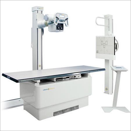
Today’s diagnostic imaging equipment typically includes:
-
An X-ray tube that generates radiation
-
A control panel for adjusting exposure settings
-
A digital detector or flat panel imaging system
-
Shielding components to reduce unnecessary exposure
Modern digital radiography systems have replaced traditional film in many facilities, improving clarity, efficiency, and data storage.
Why X-Ray Technology Matters Today
X-ray machines remain central to healthcare infrastructure and industrial inspection processes. Their importance spans multiple sectors.
In healthcare, medical imaging technology supports:
-
Detection of bone fractures
-
Identification of infections
-
Lung and chest assessments
-
Dental evaluations
-
Early disease detection
In industry, industrial radiography equipment is used to:
-
Inspect pipelines and welds
-
Analyze structural integrity
-
Test aerospace components
-
Evaluate manufacturing defects
The global reliance on diagnostic imaging equipment has increased due to aging populations, higher chronic disease rates, and expanded preventive health screening programs. Hospitals, trauma centers, outpatient clinics, and dental facilities depend on advanced imaging systems to guide clinical decisions.
Beyond medicine, security screening systems at airports and cargo facilities also rely on X-ray scanning technology to detect concealed objects without physical inspection.
The ability to visualize internal structures without invasive procedures makes X-ray systems one of the most widely used imaging technologies worldwide.
How X-Ray Machines Work
X-rays are a form of high-energy electromagnetic radiation. Inside the X-ray tube:
-
Electrons are accelerated at high speed
-
They collide with a metal target, typically tungsten
-
The collision produces X-ray photons
-
These photons travel toward the object being examined
The interaction between radiation and tissue produces contrast differences. Digital detectors convert this radiation into electronic signals, which are processed into images using specialized imaging software.
Below is a simplified comparison of imaging types:
| Imaging Type | Radiation Used | Typical Applications |
|---|---|---|
| Digital Radiography | Yes | Bone imaging, chest scans |
| Computed Radiography | Yes | General diagnostic imaging |
| CT (Computed Tomography) | Yes | Cross-sectional body imaging |
| MRI | No | Soft tissue and neurological imaging |
| Ultrasound | No | Pregnancy and organ evaluation |
X-ray systems are often integrated with PACS (Picture Archiving and Communication Systems) for digital storage and remote access, supporting hospital imaging solutions.
Recent Developments in X-Ray Technology
Over the past year, advancements in digital radiography systems and AI-supported imaging have improved workflow and diagnostic accuracy.
In 2025, healthcare technology updates have highlighted:
-
Increased adoption of portable X-ray devices in emergency care
-
Artificial intelligence integration for image analysis support
-
Enhanced dose-reduction algorithms in new imaging equipment
-
Expanded use of cloud-based radiology platforms
AI-assisted imaging tools help radiologists identify abnormalities more efficiently by highlighting areas of concern. These systems do not replace professional interpretation but assist in improving workflow efficiency.
Manufacturers have also focused on lowering radiation exposure through improved detector sensitivity and optimized exposure control systems.
Portable and mobile X-ray units are increasingly used in intensive care settings and remote healthcare environments, allowing bedside imaging without patient transport.
Regulatory Standards and Radiation Safety
Because X-rays involve ionizing radiation, their use is strictly regulated.
In the United States, regulatory oversight is shared by:
-
U.S. Food and Drug Administration
-
Environmental Protection Agency
-
State health departments
The FDA regulates diagnostic imaging equipment under the Federal Food, Drug, and Cosmetic Act. Facilities must comply with radiation safety regulations, equipment performance standards, and quality assurance protocols.
Internationally, guidance is provided by:
-
World Health Organization
-
International Atomic Energy Agency
Common compliance requirements include:
-
Regular equipment inspection
-
Radiation dose monitoring
-
Protective shielding
-
Operator certification and training
-
Documentation and reporting
Radiation exposure is measured in millisieverts (mSv). Medical imaging follows the ALARA principle — “As Low As Reasonably Achievable” — to minimize patient exposure.
Facilities must maintain radiation safety programs and ensure staff wear monitoring badges when required.
Tools and Resources for X-Ray Systems
Several professional tools support imaging management and compliance.
Common resources include:
-
Radiation dose calculators
-
Digital image processing software
-
PACS platforms
-
Quality assurance checklists
-
Equipment calibration logs
Healthcare professionals often use radiology reporting software to document findings accurately. Industry inspection teams rely on non-destructive testing (NDT) analysis tools to interpret radiographic images of materials.
Educational resources from organizations such as the Radiological Society of North America provide guidelines and continuing education for radiology professionals.
Many institutions also use structured templates to standardize imaging documentation and maintain regulatory compliance.
Safety Comparison Graph (Radiation Exposure Approximation)
Below is a simplified comparison of approximate radiation exposure levels:
| Procedure | Approximate Exposure (mSv) |
|---|---|
| Chest X-Ray | 0.1 |
| Dental X-Ray | 0.005 |
| Mammogram | 0.4 |
| CT Scan (Abdomen) | 8–10 |
| Natural Background (Year) | 3 |
These values vary depending on equipment, patient size, and imaging protocol.
Advances in advanced imaging systems continue to reduce radiation dose while maintaining image clarity.
Frequently Asked Questions
What is the difference between digital radiography and computed radiography?
Digital radiography uses flat-panel detectors that immediately convert radiation into digital images. Computed radiography uses imaging plates that require additional processing before image viewing.
Are X-rays safe?
When used appropriately and under radiation safety regulations, diagnostic X-rays are considered safe. The radiation dose is carefully controlled and minimized.
Who operates X-ray machines?
Certified radiologic technologists operate medical X-ray machines. Industrial radiography systems are handled by trained inspection professionals.
How often should imaging equipment be inspected?
Inspection frequency depends on national and regional regulations, but routine quality checks and calibration are typically required annually or semi-annually.
Can portable X-ray devices provide accurate results?
Modern portable X-ray devices are designed to produce high-quality images and are commonly used in emergency and critical care settings.
Broader Applications Beyond Medicine
While healthcare is the most recognized use, X-ray machines also play a role in:
-
Aviation security screening
-
Cargo inspection
-
Archaeological artifact analysis
-
Art restoration studies
-
Engineering failure analysis
In manufacturing, industrial radiography equipment supports infrastructure safety and compliance with engineering standards.
The versatility of X-ray technology demonstrates its impact across sectors requiring non-invasive internal visualization.
Conclusion
X-ray machines are foundational tools in modern medical imaging technology and industrial inspection. By producing internal images through controlled radiation exposure, they enable accurate diagnosis, structural evaluation, and safety assurance.
Recent innovations in digital radiography systems, AI integration, and portable X-ray devices have enhanced efficiency and reduced radiation dose. Strict radiation safety regulations and compliance standards ensure responsible use.
As healthcare systems and industrial operations continue evolving, advanced imaging systems remain central to diagnostics, safety evaluation, and technical analysis. Understanding their working principles, regulatory framework, and modern applications supports informed decision-making in clinical and technical environments.

YOU MAY ALSO LIKE