Medical Ultrasound Machines Guide: Imaging Technology and Uses
Medical ultrasound machines are diagnostic imaging systems that use high-frequency sound waves to produce real-time images of internal organs, tissues, blood vessels, and developing pregnancies.
They consist of a transducer, signal-processing electronics, imaging software, and a display. Ultrasound is widely used because it does not rely on ionizing radiation and can support a broad range of clinical examinations.
Different ultrasound systems are designed for specific medical applications, from general abdominal imaging and cardiac assessment to vascular examinations, obstetric scans, and image-guided procedures. Selecting an appropriate system requires consideration of clinical needs, image quality, workflow, safety, and equipment compatibility.
Quick Facts About Medical Ultrasound Machines
Feature | Description |
|---|---|
Primary purpose | Diagnostic medical imaging |
Imaging principle | High-frequency sound waves and returning echoes |
Main imaging modes | B-mode, M-mode, Doppler, and specialized imaging modes |
Main components | Transducer, processing unit, display, controls, and software |
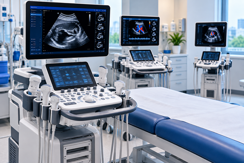
Common system formats | Cart-based, portable, handheld, and specialized systems |
Major clinical applications | Abdominal, cardiac, obstetric, vascular, and musculoskeletal imaging |
Radiation type | Uses sound waves rather than ionizing radiation |
Image output | Real-time images and selected motion or blood-flow measurements |
Key selection factors | Clinical application, transducer options, image quality, and workflow |
Main safety consideration | Appropriate acoustic output and examination duration |
What Are Medical Ultrasound Machines?
Medical ultrasound machines are electronic devices that create images by transmitting sound waves into the body and analyzing the echoes that return from tissues and organs. The system converts these echoes into visual information that clinicians use to examine anatomy, observe movement, and assess selected aspects of blood flow.
The technology is also known as diagnostic sonography. Unlike X-ray imaging and computed tomography, conventional diagnostic ultrasound does not use ionizing radiation. Its capabilities depend on the transducer, operating frequency, imaging mode, software, and the body region being examined.
Modern ultrasound equipment may include advanced image processing, automated measurements, 3D or 4D imaging, and tools for guiding certain medical procedures. These features vary by system and are not available on every model.
Why Medical Ultrasound Machines Matter
Ultrasound provides clinicians with a flexible way to examine internal structures without the need for ionizing radiation. Its ability to display movement in real time makes it particularly useful for evaluating cardiac activity, fetal movement, blood flow, and the behavior of certain tissues during examination.
Medical ultrasound also supports point-of-care assessment, in which imaging is performed close to where a patient receives care. Depending on the clinical question, this can help clinicians decide whether additional investigations or treatment are needed.
Important uses include:
Examining abdominal organs and selected soft tissues.
Monitoring pregnancy and fetal development.
Assessing cardiac structure and function.
Evaluating blood vessels and blood-flow patterns.
Investigating muscles, tendons, ligaments, and joints.
Guiding needle placement during selected procedures.
Supporting bedside assessment in emergency and critical care settings.
Ultrasound findings must be interpreted in the context of the patient's symptoms, medical history, and other clinical information. Not every condition can be reliably diagnosed using ultrasound alone.
Understanding Medical Ultrasound Imaging Technology
Simple Explanation of Ultrasound Imaging
An ultrasound transducer contains piezoelectric elements that convert electrical energy into sound pulses and returning echoes into electrical signals. The system analyzes the timing, strength, and characteristics of these echoes to create an image.
Different tissues reflect sound differently. The machine processes these variations into visual patterns that help distinguish structures such as fluid-filled spaces, soft tissues, and organ boundaries. Movement can also be assessed by tracking changes in echoes or analyzing frequency shifts associated with moving structures.
Frequency, Resolution, and Imaging Depth
Ultrasound frequency affects how much anatomical detail can be displayed and how deeply sound can penetrate. Higher-frequency transducers generally provide better spatial resolution for superficial structures, while lower-frequency transducers generally support imaging at greater depths.
This creates a practical trade-off when selecting a medical ultrasound machine. A probe suitable for thyroid or musculoskeletal imaging may not provide the penetration needed for a deep abdominal examination. Clinical systems often support multiple transducer types to accommodate different examination requirements.
How Medical Ultrasound Machines Work
Step 1: Transmitting Sound Pulses
The operator selects a transducer and examination preset appropriate for the body region. The system sends controlled electrical pulses to the transducer, which converts them into high-frequency acoustic waves.
Step 2: Receiving Returning Echoes
As the sound waves travel through the body, boundaries between tissues reflect some of the acoustic energy. The transducer receives the returning echoes and converts them into electrical signals.
Step 3: Calculating Echo Position
The system estimates the depth of reflecting structures using the time taken for echoes to return. Echo strength and other signal characteristics help determine how image information is represented.
Step 4: Processing the Image
Digital processing converts the received signals into image data. Depending on the selected mode, the system may display a two-dimensional anatomical image, movement over time, blood-flow information, or a three-dimensional representation.
Step 5: Displaying and Recording Results
The processed image appears on the monitor for real-time interpretation. Authorized users can save still images, cine loops, measurements, and examination records within the system or connected clinical infrastructure.
Step 6: Reviewing Clinical Findings
A qualified healthcare professional interprets the images in relation to the clinical question. Image quality, examination technique, and patient anatomy can affect what is visible, so findings may need correlation with other investigations.
Medical Ultrasound Imaging Workflow Table
Stage | Main Function | Output |
|---|---|---|
Examination setup | Select the appropriate probe and preset | Configured imaging system |
Pulse transmission | Send acoustic energy into the body | Sound waves |
Echo reception | Detect reflected acoustic signals | Electrical signals |
Signal processing | Estimate structure location and echo characteristics | Processed image data |
Image formation | Display anatomy or motion | Real-time image |
Measurement | Assess selected structures or flow variables | Clinical measurements |
Documentation | Save images and examination details | Stored clinical record |
Interpretation | Evaluate findings in context | Diagnostic assessment |
Main Components of Medical Ultrasound Machines
Ultrasound Transducer
The transducer is the component placed against the patient's skin or used internally for selected examinations. It transmits sound waves and receives echoes. Its shape, frequency range, and design determine which anatomical regions it can examine effectively.
Transmitter and Receiver Electronics
These electronics generate the pulses sent to the transducer and amplify the returning signals. Their performance influences the system's ability to detect useful echoes across different tissue depths.
Beamforming and Signal Processing Unit
Beamforming coordinates signals from multiple transducer elements to focus and steer the ultrasound beam. Signal processing then improves the representation of returning echoes and prepares information for image formation.
Display and Operator Controls
The monitor displays images, measurements, and system information. The control panel or touchscreen allows trained users to adjust imaging depth, gain, focus, Doppler settings, and other examination parameters.
Imaging and Measurement Functions
Imaging software provides examination presets, measurement tools, annotations, and specialized modes. Depending on the system, it may also include automated calculations or decision-support features.
Data Storage and Connectivity
Medical ultrasound machines may store examinations locally and exchange images or reports with hospital information systems and picture archiving and communication systems. Integration capabilities depend on the device, network, and clinical IT environment.
Main Components of Medical Ultrasound Machines Table
Component | Primary Function | Selection Consideration |
|---|---|---|
Transducer | Transmit and receive sound | Frequency, footprint, and examination type |
Transmitter | Generate acoustic pulses through the probe | Available operating modes |
Receiver electronics | Detect and amplify echoes | Signal sensitivity |
Beamformer | Coordinate and focus ultrasound signals | Imaging performance |
Processing unit | Convert signals into usable images | Processing capabilities |
Display | Present images and measurements | Resolution, size, and viewing ergonomics |
Operator interface | Control examination settings | Ease of use and workflow |
Storage and connectivity | Save and transfer clinical data | Compatibility and data security |
Types of Medical Ultrasound Machines
Medical ultrasound equipment can be classified by its clinical application, physical configuration, or imaging capability. These categories overlap: for example, a portable cardiac ultrasound machine may provide Doppler imaging and may also support three-dimensional cardiac examinations.
General Imaging Ultrasound Machines
General imaging systems are designed for examinations of abdominal organs, pelvic structures, superficial tissues, and other anatomical regions. They commonly support multiple transducer types and several imaging modes.
Advantages:
Flexible across multiple examination categories.
Suitable for general radiology and diagnostic imaging departments.
Often supports a broad range of measurements and presets.
Obstetric and Gynecological Ultrasound Machines
These systems support pregnancy assessment and imaging of the uterus, ovaries, and other pelvic structures. Depending on the configuration, they may provide specialized obstetric measurements, fetal imaging, and three-dimensional or four-dimensional visualization.
Advantages:
Designed around obstetric and pelvic imaging workflows.
Supports measurements used in clinical assessment.
May include specialized fetal imaging tools.
Cardiac Ultrasound Machines
Also known as echocardiography systems, cardiac ultrasound machines are designed to assess the heart's structures and movement. They commonly include M-mode, Doppler, and other tools for examining cardiac function and blood flow.
Advantages:
Specialized cardiac imaging and measurement capabilities.
Supports real-time evaluation of moving structures.
Designed for cardiac examination workflows.
Vascular Ultrasound Machines
Vascular systems are used to examine arteries and veins, including blood-flow direction and selected flow characteristics. Duplex imaging combines anatomical ultrasound with Doppler assessment, while color Doppler presents flow information overlaid on the anatomical image.
Advantages:
Supports assessment of blood vessels and circulation.
Provides anatomical and flow information.
Useful for selected vascular diagnostic procedures.
Musculoskeletal Ultrasound Machines
Musculoskeletal ultrasound focuses on muscles, tendons, ligaments, joints, and superficial soft tissues. High-frequency linear transducers are commonly used when detailed imaging of structures close to the skin is required.
Advantages:
Real-time assessment of moving soft tissues.
Supports selected image-guided procedures.
Can be useful for examining superficial structures dynamically.
Point-of-Care Ultrasound Systems
Point-of-care ultrasound, or POCUS, refers to ultrasound examinations performed by appropriately trained clinicians at the location of patient care. Systems may be cart-based, portable, or handheld, depending on the clinical environment.
Advantages:
Supports bedside assessment.
Can be deployed in emergency and critical care environments.
May streamline selected clinical decisions when appropriate expertise is available.
Handheld Ultrasound Machines
Handheld ultrasound devices are compact systems that connect to a mobile device or use an integrated display. They are designed for portability and focused examinations, although capabilities differ substantially across models.
Advantages:
Easy to transport between clinical locations.
Useful for selected bedside assessments.
Requires less physical space than many cart-based systems.
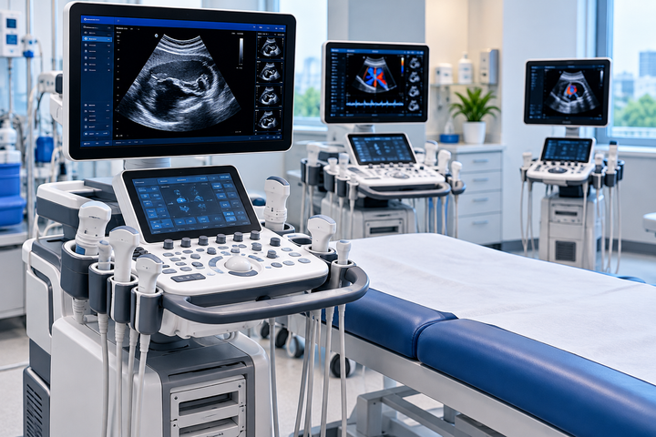
Portable and Cart-Based Ultrasound Machines
Portable systems are designed for movement between examination areas, while cart-based systems generally provide a larger console, display, and broader integration options. Neither format guarantees superior image quality; actual performance depends on the transducer, imaging technology, and intended application.
Advantages:
Portable systems support flexible deployment.
Cart-based systems may offer more extensive controls and peripherals.
Both formats can support professional diagnostic imaging when appropriately configured.
Medical Ultrasound Machine Type Comparison
System Type | Main Application | Typical Configuration | Important Capability |
|---|---|---|---|
General imaging | Abdominal and pelvic examinations | Portable or cart-based | Multiple probe support |
Obstetric and gynecological | Pregnancy and pelvic imaging | Portable or cart-based | Specialized measurements |
Cardiac | Heart structure and function | Portable or cart-based | Echocardiography and Doppler |
Vascular | Arteries and veins | Portable or cart-based | Duplex and Doppler imaging |
Musculoskeletal | Muscles, tendons, and joints | Portable or cart-based | Superficial imaging |
Point-of-care | Bedside clinical assessment | Handheld, portable, or cart-based | Rapid focused examinations |
Handheld | Selected focused examinations | Compact device | Mobility and convenient deployment |
Ultrasound Imaging Modes and Specialized Features
Two-Dimensional B-Mode Imaging
B-mode, or brightness-mode imaging, produces the familiar grayscale ultrasound image. Echo strength determines pixel brightness, allowing clinicians to assess anatomical boundaries, tissue patterns, and selected abnormalities.
It is the foundation of many abdominal, obstetric, vascular, and musculoskeletal examinations.
M-Mode Imaging
Motion mode displays the movement of structures along a selected ultrasound line over time. It is particularly associated with cardiac imaging, where clinicians can examine the motion of heart structures during the cardiac cycle.
Doppler Ultrasound
Doppler imaging uses changes in the frequency of returning echoes to assess movement, particularly blood flow. Depending on the system and examination, it may provide information about flow direction, relative velocity, and patterns of circulation.
Common Doppler modes include:
Color Doppler: Overlays color-coded flow information on an anatomical image.
Spectral Doppler: Displays a graph of Doppler frequency shifts over time.
Power Doppler: Displays the strength of Doppler signals and can be sensitive to flow in small vessels, but generally does not provide flow direction.
Three-Dimensional and Four-Dimensional Imaging
Three-dimensional ultrasound reconstructs a volume from acquired image data. Four-dimensional ultrasound displays three-dimensional information dynamically over time.
These capabilities are used in selected obstetric, gynecological, and other specialized examinations. Image quality depends on the equipment, acquisition technique, patient anatomy, and clinical application.
Contrast-Enhanced Ultrasound
Contrast-enhanced ultrasound uses approved ultrasound contrast agents to improve visualization of blood flow and selected tissue characteristics. Availability and approved applications vary by country, device, and agent.
It requires appropriate clinical assessment because contrast-enhanced examinations have different considerations from routine ultrasound imaging.
Elastography
Ultrasound elastography assesses tissue stiffness or deformation using specialized techniques. It can support selected evaluations of conditions affecting organs such as the liver and can be used in other specialized clinical applications.
Results depend on the method, examination technique, and clinical context; stiffness measurements should not be interpreted as a diagnosis in isolation.
Medical Ultrasound Machines vs. CT, MRI, and X-Ray Imaging
Medical ultrasound is one of several diagnostic imaging technologies. Each modality provides different information, so the appropriate examination depends on the clinical question, the body region, and the patient's circumstances.
Feature | Ultrasound | CT | MRI | X-Ray |
|---|---|---|---|---|
Imaging energy | Sound waves | X-rays | Magnetic fields and radiofrequency energy | X-rays |
Ionizing radiation | No | Yes | No | Yes |
Real-time imaging | Common | Available in selected applications | Available in specialized sequences | Limited in routine imaging |
Soft-tissue assessment | Useful in many accessible regions | Useful for many anatomical and emergency assessments | Excellent for many soft-tissue applications | More limited |
Blood-flow assessment | Doppler capabilities | Requires suitable CT techniques | Requires specialized techniques | Not a primary capability |
Bone evaluation | Limited for structures behind bone | Strong for many bone and trauma assessments | Useful for marrow and surrounding soft tissues | Commonly used for initial bone imaging |
Practical considerations | Probe access and operator technique | Radiation exposure and contrast considerations where applicable | Longer examinations and implant compatibility considerations | Radiation exposure and limited soft-tissue contrast |
Ultrasound is not a universal substitute for CT, MRI, or X-ray imaging. For example, air and bone can obstruct sound transmission, while some deep structures may be difficult to visualize. Clinicians choose the modality that best addresses the diagnostic question.
Requirements for Medical Ultrasound Equipment
Transducer Compatibility
The transducer must match the intended examination. Linear probes are commonly used for superficial structures, curved-array probes for many abdominal examinations, and phased-array probes for cardiac imaging and selected examinations through narrow acoustic windows.
Compatibility must be confirmed for the specific ultrasound console and transducer model.
Clinical Workflow and Ergonomics
A medical ultrasound machine should fit the examination environment, expected patient volume, and movement requirements. Considerations include monitor visibility, adjustable controls, probe storage, mobility, battery operation where available, and the ease of changing examination presets.
Ergonomics matter because repeated scanning can place strain on clinicians. Device placement and workflow design should support safe, comfortable use.
Connectivity and Data Management
Facilities may require Digital Imaging and Communications in Medicine compatibility, often called DICOM, to exchange images with clinical imaging systems. Integration with electronic health records, worklists, and picture archiving systems can reduce manual data entry and improve record management.
Connectivity should be evaluated alongside access controls, secure data transfer, software support, and the facility's privacy requirements.
Regulatory and Clinical Suitability
Medical ultrasound equipment should be authorized or registered for its intended use under the applicable jurisdiction's regulatory framework. Procurement teams should confirm that the selected system supports the required examinations and that clinicians have appropriate training.
Technical specifications alone do not establish clinical suitability. Image quality, examination protocols, maintenance support, and quality assurance also need consideration.
Benefits and Advantages of Medical Ultrasound Machines
Medical ultrasound systems offer several practical advantages when used for appropriate clinical indications.
No ionizing radiation: Diagnostic ultrasound uses acoustic energy rather than X-rays.
Real-time visualization: Supports assessment of movement and selected physiological processes.
Broad clinical utility: Used in abdominal, obstetric, cardiac, vascular, and musculoskeletal examinations.
Portability: Handheld and portable systems support selected bedside and remote-location examinations.
Image-guided procedures: Helps clinicians visualize anatomy during certain needle-guided and interventional procedures.
Repeat examination capability: Can support follow-up imaging when clinically appropriate.
Flexible examination formats: Different transducers and imaging modes support a range of clinical needs.
Potential workflow efficiency: Bedside imaging may reduce the need to move certain patients for selected assessments.
These advantages do not mean ultrasound is always the safest or most informative choice for every condition. Appropriate use requires a clinical indication, trained personnel, and consideration of alternative diagnostic methods.
Industry Applications of Medical Ultrasound Machines
Hospitals and Radiology Departments
Hospitals use diagnostic ultrasound for abdominal imaging, pelvic examinations, vascular assessment, and many other clinical investigations. General imaging systems may serve several departments, while specialized units support focused examinations.
Obstetrics and Gynecology
Ultrasound supports assessment of pregnancy, fetal development, the uterus, ovaries, and other pelvic structures. Examination protocols and the choice of imaging mode depend on the clinical indication and stage of care.
Cardiology
Echocardiography uses ultrasound to assess heart chambers, valves, movement, and selected measures of cardiac function. Doppler techniques provide information about blood flow through the heart and major vessels.
Emergency and Critical Care
Point-of-care ultrasound can assist trained clinicians with focused assessments in emergency departments, intensive care units, and other acute-care environments. Findings may help guide immediate clinical decisions but must be interpreted within the broader patient assessment.
Vascular Medicine
Vascular ultrasound helps assess selected conditions affecting arteries and veins. Duplex imaging combines anatomical information with Doppler measurements to evaluate blood-flow characteristics.
Musculoskeletal Medicine
Clinicians use ultrasound to examine superficial muscles, tendons, ligaments, and joints. It can also guide selected injections and other procedures when appropriate.
Outpatient Clinics and Primary Care
Portable ultrasound systems may support focused examinations in clinics where the equipment, clinician expertise, and clinical protocols are suitable. The scope of use depends on training, device capabilities, and local practice requirements.
Medical Ultrasound Machine Applications Table
Clinical Setting | Common Examination | Relevant Technology |
|---|---|---|
Radiology | Abdominal and pelvic imaging | B-mode and specialized modes |
Obstetrics | Fetal and pregnancy assessment | B-mode, Doppler when indicated, 3D/4D where appropriate |
Cardiology | Heart structure and function | Echocardiography and Doppler |
Emergency care | Focused bedside assessment | Portable or handheld ultrasound |
Vascular medicine | Arterial and venous evaluation | Duplex and Doppler |
Musculoskeletal care | Tendon, muscle, and joint imaging | High-frequency linear probes |
Outpatient clinics | Focused diagnostic assessment | Portable systems suited to the examination |
Image-guided procedures | Needle and instrument visualization | Real-time ultrasound guidance |
Medical Ultrasound Standards and Patient Safety
Medical ultrasound equipment must meet applicable medical-device requirements, and clinical use should follow established safety practices. Standards, regulatory authorization, and personnel requirements differ across countries, so healthcare facilities should confirm the rules that apply to their location and intended use.
IEC 60601-2-37
IEC 60601-2-37:2024 specifies particular requirements for the basic safety and essential performance of ultrasonic medical diagnostic and monitoring equipment. It is relevant to equipment design and safety evaluation, alongside applicable general medical electrical equipment standards.
FDA Medical Device Requirements
In the United States, diagnostic ultrasound systems and transducers are subject to applicable Food and Drug Administration medical-device requirements. Regulatory status should be checked for the particular device and its intended use.
Other countries maintain their own medical-device authorization or conformity assessment frameworks.
ALARA Principle
ALARA means “as low as reasonably achievable.” It guides clinicians to use acoustic output and examination duration appropriate to the diagnostic purpose while obtaining the necessary information.
Diagnostic ultrasound does not use ionizing radiation, but acoustic energy can produce thermal and mechanical effects in tissue. It should therefore be used prudently by appropriately trained healthcare professionals.
Infection Prevention and Probe Hygiene
Transducers must be cleaned and disinfected according to their intended use and the manufacturer's instructions. External probes, internal-use probes, and equipment used during sterile procedures may require different levels of reprocessing.
Compatible coupling gel, protective covers where indicated, and correct handling procedures help reduce contamination risks. A probe should not be returned to clinical use until the required cleaning and disinfection process has been completed.
Clinical Quality Assurance
Quality assurance helps confirm that ultrasound systems produce images suitable for their intended clinical applications. Programs may include equipment inspections, image-quality tests, transducer assessments, documentation, and corrective action when performance problems are identified.
A qualified medical physicist, biomedical engineering professional, or appropriately trained service specialist may be required, depending on the task and applicable rules.
Common Medical Ultrasound Machine Challenges and Solutions
Challenge | Possible Cause | Appropriate Response |
|---|---|---|
Poor image clarity | Incorrect settings, unsuitable probe, or difficult acoustic access | Review the examination setup and transducer selection |
Limited imaging depth | Frequency too high or inadequate acoustic penetration | Adjust settings and select a suitable probe |
Image artifacts | Acoustic interactions, motion, or equipment issues | Assess the artifact and verify findings using appropriate techniques |
Inconsistent measurements | Different acquisition methods or operator technique | Follow standardized examination protocols |
Transducer damage | Wear, impact, or improper handling | Remove damaged equipment from use and arrange assessment |
Connectivity problems | Network configuration or compatibility issues | Review system configuration and obtain technical support |
Inadequate data protection | Weak access controls or insecure data transfer | Apply facility-approved information security controls |
Infection-control concerns | Incomplete cleaning or incorrect disinfection | Follow the approved reprocessing protocol |
Equipment downtime | Hardware faults or overdue maintenance | Arrange qualified technical assessment |
Uncertain clinical findings | Limited visualization or nonspecific appearances | Seek appropriate clinical review or alternative imaging |
How to Choose a Medical Ultrasound Machine
Selecting a medical ultrasound machine requires matching its capabilities to the examinations performed by the facility. A handheld device designed for focused bedside assessment may not meet the needs of a high-volume radiology department, while a specialized cardiac system may not be the most suitable choice for a general imaging practice.
The evaluation should consider clinical performance, probe compatibility, ergonomics, data integration, regulatory status, and long-term equipment support.
Selection Factor | What to Evaluate | Why It Matters |
|---|---|---|
Clinical specialty | General imaging, cardiac, obstetric, vascular, or musculoskeletal use | Determines required imaging capabilities |
Transducer selection | Probe types, frequency ranges, and compatibility | Affects access, depth, and resolution |
Imaging modes | B-mode, M-mode, Doppler, 3D/4D, or specialized functions | Determines the examinations the system can support |
Image quality | Resolution, penetration, sensitivity, and artifact handling | Influences diagnostic usefulness |
Portability | Handheld, portable, or cart-based format | Must fit the clinical environment |
Ergonomics | Display position, controls, and probe handling | Supports efficient and comfortable scanning |
Data integration | DICOM, worklists, and record-system compatibility | Supports clinical documentation |
Safety and compliance | Regulatory status and applicable standards | Helps establish suitability for intended use |
Quality assurance | Testing procedures and technical support | Supports ongoing image quality |
Maintenance | Warranty terms, replacement probes, and repair support | Affects continuity of clinical operation |
Training | User training and application support | Helps staff use the equipment appropriately |
Expansion needs | Additional probes, software features, and connectivity | Supports changing clinical requirements |
The most appropriate system is the one that meets the facility's clinical requirements and operating constraints, not necessarily the device with the greatest number of features.
Maintenance Best Practices for Medical Ultrasound Machines
Routine maintenance helps preserve image quality, electrical safety, and dependable operation. The maintenance schedule should follow the manufacturer's instructions, facility policy, and applicable regulatory requirements.
Inspect Transducers and Cables
Check probe surfaces, cable insulation, connectors, and strain-relief points for visible damage. Damaged transducers may produce artifacts, compromise electrical safety, or affect clinical performance.
Clean and Disinfect Equipment Correctly
Use cleaning agents and disinfectants compatible with the specific probe and console. Follow the required reprocessing procedure for the intended use, including any applicable high-level disinfection requirements for internal-use transducers.
Verify Image Quality
Quality-control procedures can identify changes in image uniformity, sensitivity, resolution, or the presence of artifacts. Testing should use suitable procedures and equipment, with qualified personnel interpreting results.
Maintain Software and System Configuration
Software updates, security changes, and configuration modifications should follow approved facility procedures. Changes that affect clinical functions should be assessed and documented before the equipment is returned to routine use.
Review Electrical and Mechanical Safety
Inspect power connections, wheels, brakes, displays, controls, and other relevant components according to the maintenance plan. Repairs and electrical safety testing should be performed by qualified personnel when required.
Keep Maintenance Records
Document equipment inspections, faults, repairs, quality-control results, and relevant software changes. Records help identify recurring problems and provide evidence that the equipment is maintained according to the facility's requirements.
Limitations of Medical Ultrasound Machines
Although ultrasound is versatile, its diagnostic capabilities are constrained by the physical behavior of sound waves and the conditions under which images are acquired.
Bone obstruction: Bone reflects and attenuates sound, limiting views of structures behind it.
Air interference: Gas can interfere with acoustic transmission, making some areas difficult to examine.
Patient-dependent image quality: Body composition, anatomy, positioning, and other factors can affect visualization.
Operator dependence: Examination technique and experience influence image acquisition and interpretation.
Limited field of view: Ultrasound examines a selected region rather than routinely producing a comprehensive whole-body image.
Specialized expertise requirements: Some applications require advanced training and specialized equipment.
Not a replacement for every imaging method: CT, MRI, X-ray, or other investigations may be more suitable for specific diagnostic questions.
Ongoing safety and hygiene obligations: Proper acoustic exposure, equipment maintenance, and probe reprocessing remain necessary.
A normal or inconclusive ultrasound result does not necessarily exclude disease. Further clinical assessment or additional imaging may be appropriate when the findings do not resolve the diagnostic question.
Future Trends in Medical Ultrasound Technology
Artificial Intelligence-Assisted Imaging
AI-enabled ultrasound features can help with image acquisition, anatomical identification, measurements, and workflow consistency. Their availability and validated clinical applications vary by device. AI-generated outputs require appropriate clinical oversight and should not be treated as universally reliable diagnoses.
Portable and Handheld Systems
Compact ultrasound devices are expanding access to focused imaging in bedside, outpatient, emergency, and other clinical settings. Their practical value depends on image quality, intended use, probe options, battery performance, and the expertise of the operator.
Automated Measurements
Automated measurement tools can assist with selected cardiac, obstetric, and general imaging tasks. These features may reduce repetitive manual work, although results still require review for accuracy and clinical relevance.
Advanced Image Reconstruction
Developments in signal processing and image reconstruction aim to improve image clarity, reduce certain artifacts, and support visualization under challenging examination conditions. Performance gains depend on the clinical application and system implementation.
Connected Clinical Workflows
Improved integration with imaging archives and electronic health records can streamline documentation and examination management. Secure connectivity, data governance, and compatibility remain essential as ultrasound systems become more integrated with hospital infrastructure.
Frequently Asked Questions About Medical Ultrasound Machines
1. What is a medical ultrasound machine used for?
A medical ultrasound machine creates images of internal structures using high-frequency sound waves. It is used for examinations involving organs, pregnancy, the heart, blood vessels, muscles, tendons, and other tissues.
2. How does a diagnostic ultrasound machine create images?
A transducer sends sound pulses into the body and receives returning echoes. The system processes the echoes to estimate the location and characteristics of tissue boundaries and displays the results as images.
3. What are the main types of medical ultrasound machines?
Common categories include general imaging, obstetric and gynecological, cardiac, vascular, musculoskeletal, and point-of-care systems. Equipment can also be classified as handheld, portable, or cart-based, depending on its physical configuration.
4. What is the difference between 2D, 3D, and 4D ultrasound?
Two-dimensional ultrasound displays cross-sectional grayscale images. Three-dimensional ultrasound reconstructs a volume, while four-dimensional ultrasound displays three-dimensional information dynamically over time.
5. What is Doppler ultrasound used for?
Doppler ultrasound evaluates movement, particularly blood flow. Depending on the mode, it can display flow direction, relative velocity, and patterns that help clinicians assess circulation.
6. Is medical ultrasound safe?
Diagnostic ultrasound does not use ionizing radiation and has a well-established role in medical imaging. However, acoustic energy can produce biological effects under certain conditions, so examinations should be clinically justified and performed prudently by trained professionals.
7. How often should ultrasound machines be maintained?
Maintenance frequency depends on the equipment, manufacturer instructions, clinical use, and applicable quality-assurance requirements. Facilities should establish documented inspection, performance testing, cleaning, and technical maintenance procedures.
8. What factors affect ultrasound image quality?
Image quality depends on transducer frequency, acoustic penetration, system settings, signal processing, patient anatomy, and operator technique. Selecting the appropriate probe and examination settings helps optimize the image for the clinical question.
9. Can handheld ultrasound machines replace cart-based systems?
Handheld devices can support many focused examinations, but their capabilities vary. Cart-based systems may provide a broader selection of probes, imaging modes, controls, and specialized clinical features, making them more appropriate for certain diagnostic departments.
10. What standards apply to medical ultrasound equipment?
IEC 60601-2-37 addresses the basic safety and essential performance of ultrasonic medical diagnostic and monitoring equipment. Additional medical-device regulations, infection-control requirements, clinical quality-assurance procedures, and personnel requirements depend on the jurisdiction and intended use.
Conclusion
Medical ultrasound machines provide versatile diagnostic imaging through sound-wave transmission, echo processing, and real-time image formation. Their applications range from general and obstetric imaging to cardiac, vascular, and point-of-care examinations. Choosing suitable equipment requires attention to clinical needs, transducer compatibility, image quality, safety, connectivity, and maintenance. Appropriate training, quality assurance, and prudent clinical use help ensure that ultrasound technology supports reliable patient assessment.