Learn About Dental CBCT Scanners: Discover Information, Insights, Tips, Advice, and Useful Resources
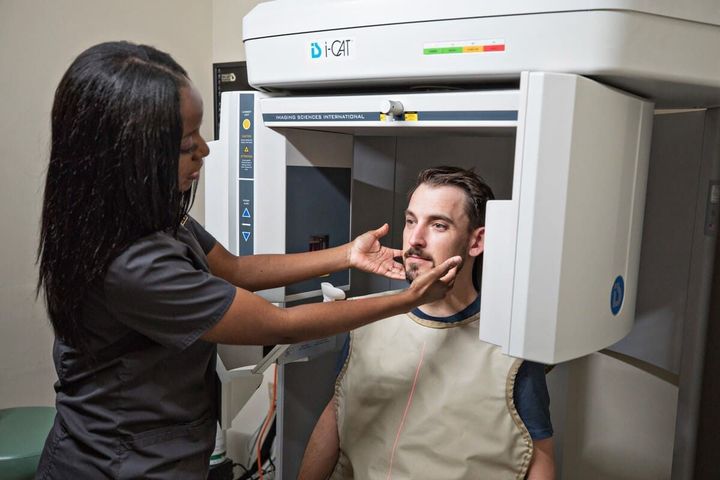
Dental CBCT scanners, also known as Cone Beam Computed Tomography scanners, are advanced imaging systems used in dentistry to create detailed three-dimensional images of teeth, jaws, facial bones, nerves, and surrounding structures. Unlike traditional dental radiography, CBCT technology captures multiple images from different angles and combines them into a comprehensive 3D view.
The development of dental CBCT technology was driven by the need for more accurate diagnostic information in oral healthcare. Traditional two-dimensional imaging can sometimes make it difficult to evaluate complex anatomical structures. CBCT scanners help dental professionals visualize areas that may not be clearly visible using conventional imaging methods.
Today, CBCT imaging is commonly used for dental implant planning, orthodontic assessments, oral surgery evaluations, airway analysis, temporomandibular joint examinations, and endodontic procedures. The technology supports improved visualization of bone structures and anatomical relationships.
Many modern systems incorporate advanced imaging software, digital workflow integration, artificial intelligence-assisted analysis, and cloud-based data management. As digital dentistry continues to evolve, CBCT scanners have become an important component of diagnostic imaging.
Why Dental CBCT Scanners Matter
The growing use of digital dental imaging has increased the importance of CBCT scanners across many areas of oral healthcare. Accurate imaging helps clinicians better understand patient anatomy and supports informed treatment planning.
Several factors contribute to the significance of CBCT technology:
- Enhanced visualization of teeth and bone structures
- Improved assessment of impacted teeth
- Better understanding of nerve pathways
- Support for orthodontic planning
- Assistance with implant positioning analysis
- Evaluation of jaw disorders and facial structures
- Integration with digital dentistry workflows
The technology affects multiple groups, including:
| Stakeholder | Potential Benefits |
|---|---|
| Dental Professionals | Detailed diagnostic information |
| Orthodontists | Improved treatment planning |
| Oral Surgeons | Enhanced anatomical visualization |
| Endodontists | Better root canal assessment |
| Patients | More informed clinical evaluations |
| Researchers | Access to detailed imaging datasets |
Dental CBCT scanners also help address challenges associated with complex dental anatomy. Three-dimensional imaging can reveal structures that may overlap on traditional radiographs, supporting more comprehensive evaluations.
Recent Updates and Industry Trends
Dental imaging technology continues to advance rapidly. During 2025 and 2026, several notable trends have influenced the CBCT scanner market and broader digital dentistry ecosystem.
Artificial Intelligence Integration
Many imaging software platforms now include AI-powered tools that assist with image interpretation, anatomical identification, airway analysis, and treatment planning support. These systems are designed to improve workflow efficiency and enhance image review processes.
Lower Radiation Optimization
Manufacturers continue to focus on radiation dose optimization. New imaging protocols aim to provide diagnostic-quality images while minimizing patient exposure through improved sensors and image reconstruction algorithms.
Cloud-Based Imaging Platforms
Cloud connectivity has become increasingly common. Imaging data can be securely stored, shared, and reviewed across multiple locations, supporting collaborative treatment planning and remote consultations.
Digital Workflow Expansion
CBCT scanners are increasingly integrated with:
- Intraoral scanners
- Digital treatment planning software
- Surgical guide design systems
- Orthodontic simulation platforms
- Digital laboratory workflows
Enhanced Image Resolution
Recent advancements have improved image clarity for specific diagnostic applications. Improved voxel resolution and image-processing technologies support detailed visualization of anatomical structures.
The chart below summarizes major technology trends observed across the digital dental imaging sector.
| Trend | Industry Focus |
| Artificial Intelligence | Image analysis support |
| Cloud Integration | Data accessibility |
| Digital Workflows | Connected dentistry |
| Dose Optimization | Radiation management |
| Advanced Software | Diagnostic efficiency |
| High-Resolution Imaging | Improved visualization |
These developments reflect broader efforts to enhance diagnostic capabilities while supporting efficient clinical workflows.
Regulations, Standards, and Policies
Dental CBCT scanners are subject to various regulations and professional guidelines designed to ensure patient safety, image quality, and responsible use.
Healthcare authorities and regulatory agencies generally require imaging equipment to meet established standards related to radiation safety, device performance, and quality assurance.
Common regulatory considerations include:
- Radiation protection requirements
- Equipment certification standards
- Quality control procedures
- Operator training recommendations
- Patient imaging justification principles
- Data privacy and record management requirements
Many countries follow guidance based on recommendations from organizations such as:
- International Atomic Energy Agency
- International Commission on Radiological Protection
- World Health Organization
Healthcare facilities are often encouraged to apply the ALARA principle, which stands for "As Low As Reasonably Achievable." This approach emphasizes minimizing radiation exposure while obtaining clinically useful diagnostic information.
Data protection regulations may also affect CBCT imaging systems because patient images are considered healthcare records. Secure storage, controlled access, and appropriate data management practices are important components of compliance.
As digital dentistry expands, policymakers continue evaluating standards related to artificial intelligence, cloud-based healthcare systems, and digital medical records.
Helpful Tools and Resources
A variety of tools and educational resources can help individuals learn more about dental CBCT technology and digital imaging.
Useful categories include:
Educational Resources
- Academic dental journals
- Dental radiology textbooks
- Continuing education programs
- Professional association publications
- University research libraries
Imaging Software Platforms
- 3D image viewers
- Treatment planning applications
- Implant planning software
- Airway analysis tools
- Surgical simulation platforms
Research Databases
- PubMed
- National Center for Biotechnology Information
- Cochrane Collaboration
Radiation Safety Resources
- Radiation protection guidelines
- Quality assurance checklists
- Imaging protocol references
- Educational webinars
Digital Workflow Resources
- 3D model visualization tools
- DICOM file viewers
- Imaging data management platforms
- Clinical documentation templates
The table below summarizes common resource categories.
| Resource Type | Purpose |
| Research Databases | Scientific information |
| Educational Courses | Knowledge development |
| Imaging Software | Image analysis |
| Safety Guidelines | Radiation awareness |
| Digital Workflow Tools | Treatment planning support |
| Professional Publications | Industry updates |
When reviewing information about CBCT scanners, it is helpful to rely on evidence-based resources, peer-reviewed research, and recognized professional organizations.
Frequently Asked Questions
What does CBCT stand for?
CBCT stands for Cone Beam Computed Tomography. It is a specialized imaging technology that produces three-dimensional images of dental and facial structures.
How is CBCT different from traditional dental X-rays?
Traditional dental radiographs typically create two-dimensional images. CBCT scanners generate three-dimensional views, allowing more detailed visualization of anatomical structures from multiple perspectives.
Why is 3D imaging useful in dentistry?
Three-dimensional imaging can help improve understanding of tooth position, bone anatomy, nerve pathways, jaw structures, and other complex areas that may be difficult to evaluate with conventional imaging.
Does CBCT technology use radiation?
Yes. CBCT scanners use ionizing radiation to produce images. Modern systems often incorporate dose optimization techniques and follow radiation safety principles to support appropriate imaging practices.
Can CBCT images be used with digital treatment planning software?
Yes. Many CBCT systems integrate with digital dentistry platforms, including implant planning software, orthodontic applications, surgical guide design tools, and 3D visualization programs.
Are CBCT scanners used only for dental implants?
No. While implant planning is a common application, CBCT technology is also used in orthodontics, oral surgery, endodontics, airway assessment, temporomandibular joint evaluation, and other diagnostic procedures.
Conclusion
Dental CBCT scanners represent a significant advancement in diagnostic imaging within modern dentistry. By providing detailed three-dimensional views of teeth, bone structures, and surrounding anatomy, these systems support comprehensive clinical evaluation and digital treatment planning.
Recent developments in artificial intelligence, cloud connectivity, image processing, and workflow integration continue to shape the future of CBCT technology. At the same time, regulatory standards and radiation safety guidelines remain important factors influencing responsible use.
As digital dentistry evolves, understanding the fundamentals of CBCT imaging can help individuals stay informed about one of the most important technologies supporting contemporary dental diagnostics. Continued research, education, and technological innovation are expected to further enhance imaging capabilities and improve the overall quality of dental care worldwide.