Dental X-Ray Machines Explained: Explore Guide, Tips, Insights, Knowledge, Facts, and Helpful Resources
Dental X-ray machines are specialized diagnostic imaging systems used to capture images of teeth, gums, jawbones, and surrounding oral structures. These machines help dental professionals examine areas that may not be visible during a routine visual examination.
The concept of dental radiography has existed for more than a century. Since the discovery of X-rays in 1895, imaging technology has evolved significantly. Modern dental imaging equipment now provides clearer images, faster processing, and improved radiation management compared with earlier systems.
Dental X-ray machines are commonly used during oral examinations to identify dental conditions such as tooth decay, bone loss, impacted teeth, infections, cysts, and other structural abnormalities. They play an important role in preventive dentistry and long-term oral health management.
Types of Dental X-Ray Machines
Different imaging systems are designed for specific diagnostic purposes.
| Type | Primary Purpose |
|---|---|
| Intraoral X-Ray | Captures detailed images from inside the mouth |
| Bitewing X-Ray | Detects cavities between teeth |
| Periapical X-Ray | Shows entire tooth structure and roots |
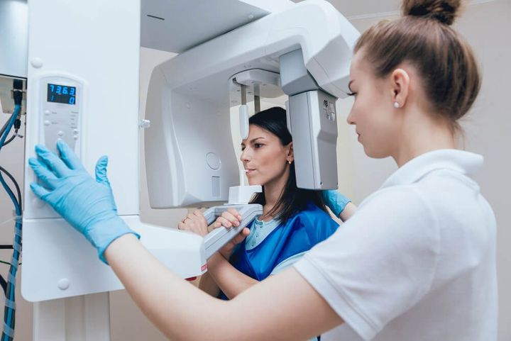
| Panoramic X-Ray | Provides a full-mouth image |
| Cephalometric X-Ray | Evaluates jaw and facial relationships |
| Cone Beam CT (CBCT) | Produces detailed 3D images of oral structures |
Digital dental imaging systems have become increasingly common because they offer enhanced image quality and efficient image management.
Why Dental X-Ray Machines Matter Today
Dental health affects overall well-being. Conditions that begin in the mouth can influence nutrition, speech, comfort, and quality of life. Dental X-ray machines help identify issues before they become more serious.
Early detection remains one of the biggest advantages of dental radiography. Small cavities, hidden infections, and jawbone changes can often be discovered before symptoms appear.
The technology benefits multiple groups:
- Patients seeking preventive dental care
- Children undergoing dental development assessments
- Adults monitoring oral health conditions
- Orthodontic patients receiving treatment planning
- Dental professionals conducting comprehensive evaluations
- Researchers studying oral health trends
Modern dental imaging also supports evidence-based treatment decisions. Detailed images allow healthcare professionals to assess conditions accurately and monitor changes over time.
Problems Dental X-Ray Machines Help Address
Dental imaging can assist in identifying:
- Tooth decay between teeth
- Gum disease-related bone loss
- Impacted wisdom teeth
- Root infections
- Dental trauma and fractures
- Jaw abnormalities
- Developmental dental issues
- Oral cysts and lesions
Without diagnostic imaging, many of these conditions may remain undetected until they become more advanced.
Key Benefits of Modern Dental Imaging
| Benefit | Explanation |
|---|---|
| Improved Detection | Identifies hidden oral conditions |
| Better Treatment Planning | Supports accurate clinical decisions |
| Digital Records | Simplifies storage and retrieval |
| Enhanced Image Quality | Provides detailed visualization |
| Faster Results | Images are available quickly |
| Reduced Repeat Imaging | Improved accuracy lowers retakes |
Recent Updates and Trends in Dental X-Ray Technology
The dental imaging industry has experienced several developments during 2025 and 2026.
One notable trend is the continued expansion of digital radiography systems. Many dental practices have transitioned from traditional film-based imaging to digital platforms, improving image accessibility and workflow efficiency.
Artificial intelligence-assisted image analysis has also gained attention throughout 2025. AI-supported software tools are increasingly being evaluated to help identify potential abnormalities in dental radiographs. These technologies are designed to support clinical interpretation rather than replace professional judgment.
Another significant development has been the growing adoption of Cone Beam Computed Tomography (CBCT) systems. CBCT technology provides highly detailed three-dimensional views of oral structures, supporting advanced diagnostic assessments and treatment planning.
Cloud-based dental imaging platforms have continued to expand during 2025 and early 2026. These systems allow secure image sharing among healthcare providers and improve access to patient records.
Recent industry discussions have also focused on radiation optimization strategies. Manufacturers continue to introduce imaging systems designed to maintain diagnostic quality while minimizing radiation exposure through improved sensors and software processing.
Emerging Technologies
Current innovations include:
- AI-assisted diagnostic imaging
- Enhanced digital sensors
- Advanced CBCT visualization
- Cloud-based image management
- Integrated imaging software platforms
- Improved image reconstruction algorithms
These developments aim to enhance diagnostic efficiency and clinical accuracy.
Laws, Regulations, and Policy Considerations
Dental X-ray machines operate within regulatory frameworks designed to ensure patient safety and equipment performance.
Most countries regulate dental radiography through healthcare, radiation protection, and medical device authorities. Requirements typically include equipment inspection, operator training, maintenance standards, and radiation safety procedures.
Key regulatory areas often include:
- Radiation protection standards
- Equipment certification requirements
- Quality assurance programs
- Patient record management
- Operator education and competency
- Medical device compliance
Healthcare facilities generally follow established radiation safety principles such as justification, optimization, and dose limitation where applicable.
Common Radiation Safety Principles
| Principle | Purpose |
|---|---|
| Justification | Imaging should have a clinical reason |
| Optimization | Radiation exposure should be minimized |
| Quality Assurance | Equipment should function properly |
| Staff Training | Operators should follow safety protocols |
| Monitoring | Imaging procedures should be regularly reviewed |
Many governments and healthcare organizations periodically update guidance as imaging technologies evolve and new scientific evidence becomes available.
Helpful Tools and Resources
Several digital tools and educational resources support learning and management of dental imaging technologies.
Educational Resources
- Dental radiography learning platforms
- Oral health education portals
- Medical imaging reference libraries
- Continuing education programs
- Academic dental journals
Software and Digital Tools
- Digital dental imaging software
- CBCT visualization applications
- Image annotation platforms
- Electronic health record integration tools
- Secure cloud imaging systems
Professional Resources
- Radiation safety guidelines
- Imaging quality assurance checklists
- Clinical imaging protocols
- Dental radiography reference manuals
- Professional training modules
Useful Templates and Documentation
Organizations often use:
- Imaging consent forms
- Equipment maintenance logs
- Quality control records
- Radiation safety checklists
- Clinical documentation templates
These resources help maintain consistent imaging practices and support regulatory compliance.
Dental X-Ray Technology at a Glance
Traditional Film Imaging
          │
          ▼
Digital Radiography
          │
          ▼
Enhanced Image Processing
          │
          ▼
3D CBCT Imaging
          │
          ▼
AI-Assisted Analysis
          │
          ▼
Cloud-Based Image Management
The progression illustrates how dental imaging technology has evolved toward greater accuracy, accessibility, and digital integration.
Frequently Asked Questions
What is a dental X-ray machine used for?
A dental X-ray machine captures images of teeth, jawbones, and surrounding oral structures. These images help identify conditions that may not be visible during a standard oral examination.
Are dental X-rays safe?
Dental X-rays are generally considered safe when used appropriately and according to established radiation safety guidelines. Modern digital systems often use significantly lower radiation levels than older imaging technologies.
What is the difference between digital dental imaging and traditional film imaging?
Digital dental imaging produces electronic images that can be viewed immediately on a computer. Traditional film imaging requires chemical processing and physical storage.
What is Cone Beam CT (CBCT)?
Cone Beam Computed Tomography is an advanced imaging technology that creates detailed three-dimensional images of oral and facial structures. It is commonly used for complex diagnostic and treatment planning purposes.
How often are dental X-rays needed?
The frequency depends on individual oral health needs, age, risk factors, and professional recommendations. Imaging decisions are typically based on clinical assessment and established guidelines.
Can dental X-rays detect problems early?
Yes. Dental radiographs can reveal cavities, infections, bone changes, impacted teeth, and other conditions before noticeable symptoms develop, allowing earlier evaluation and management.
Conclusion
Dental X-ray machines remain an essential component of modern oral healthcare. These imaging systems provide valuable insights into dental and jaw structures that cannot always be observed through visual examination alone. Advances in digital dental imaging, Cone Beam CT technology, AI-assisted analysis, and cloud-based imaging platforms continue to improve diagnostic capabilities and workflow efficiency.
As dental radiography evolves, patient safety, image quality, and regulatory compliance remain important priorities. Understanding how dental X-ray machines work, their benefits, and the technologies shaping their future helps individuals appreciate the role diagnostic imaging plays in maintaining oral health and supporting informed healthcare decisions.