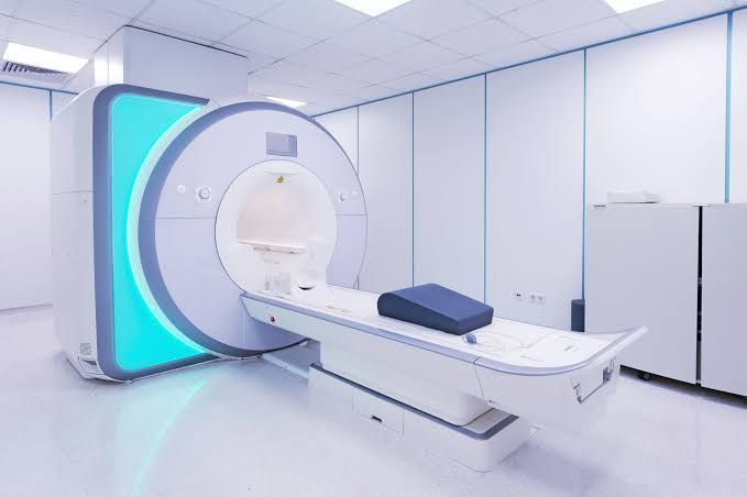
Unlike X-ray and CT imaging, MRI does not use ionizing radiation. It is widely used for imaging the brain, spine, joints, muscles, abdominal organs, blood vessels, and other soft tissues.
A hospital MRI system is more than the scanner itself. A complete MRI environment can involve the magnet, patient table, radiofrequency systems, gradient coils, computer systems, cooling infrastructure, shielding, safety equipment, electrical systems, and specialized room design.
This guide explains major MRI machine types, field strengths, important specifications, installation requirements, safety considerations, operational factors, cost considerations, and emerging MRI technologies.
What Is an MRI Machine?
An MRI machine uses a strong static magnetic field to influence the behavior of hydrogen nuclei in the body. Radiofrequency energy is then used to manipulate these nuclei, and the signals produced as they return toward equilibrium are detected and processed into images.
A typical MRI system includes:
-
Main magnet
-
Gradient coils
-
Radiofrequency coils
-
Patient table
-
RF electronics
-
Computer and reconstruction system
-
Cooling systems
-
Control console
The scanner operates as part of a larger imaging environment designed around electromagnetic safety and controlled access.
Main Components of an MRI System
Main Magnet
The magnet generates the primary static magnetic field.
MRI systems are commonly described by their magnetic-field strength, measured in tesla (T).
Common clinical configurations include:
Higher-field research and specialized systems also exist.
Gradient Coils
Gradient coils generate controlled variations in the magnetic field.
They help determine spatial information within the body and are fundamental to MRI image formation.
Gradient performance can influence:
-
Spatial resolution
-
Imaging speed
-
Diffusion imaging
-
Advanced MRI sequences
Radiofrequency Coils
RF coils transmit and/or receive radiofrequency signals.
Different coils are designed for different anatomical regions, including:
-
Head
-
Spine
-
Knee
-
Shoulder
-
Breast
-
Abdomen
-
Extremities
Coil design can significantly influence signal quality.
Patient Table
The patient table moves the person into the magnet bore.
It needs to provide:
-
Accurate positioning
-
Stability
-
Patient comfort
-
Appropriate weight capacity
-
Compatibility with imaging protocols
Computer and Reconstruction System
MRI generates large quantities of imaging data.
Computer systems process raw signals into diagnostic images that can be reviewed by radiologists and integrated into hospital imaging systems.
Types of Hospital MRI Machines
MRI systems can be classified in several ways.
1.5 Tesla MRI
1.5T systems are widely used for routine clinical imaging.
Potential advantages include:
-
Broad clinical applications
-
Established technology
-
Compatibility with many protocols
-
Extensive clinical experience
3 Tesla MRI
3T systems provide a higher magnetic-field strength than 1.5T systems.
They can offer increased signal-to-noise ratio for many applications and may support high-resolution imaging and advanced protocols.
However, higher field strength can also introduce additional technical considerations, including susceptibility effects and specific absorption-rate management.
Low-Field MRI
Lower-field systems operate at substantially lower magnetic-field strengths.
They can have applications in specific environments where system size, accessibility, or specialized imaging requirements are important.
Open MRI
Open MRI designs provide a more open physical configuration.
They may be considered for certain patients who experience difficulty with conventional enclosed systems or where accessibility is an important consideration.
Image quality and clinical capabilities depend on the specific system design.
Wide-Bore MRI
Wide-bore systems provide a larger patient opening than conventional narrow-bore configurations.
They can improve physical accessibility and patient comfort while retaining many characteristics of conventional cylindrical MRI systems.
Understanding MRI Field Strength
Field strength is one of the most commonly discussed MRI specifications.
1.5T vs 3T
The choice between 1.5T and 3T depends on the intended clinical applications, patient population, protocols, infrastructure, and imaging requirements.
3T can provide higher signal-to-noise characteristics in many circumstances, which may support higher-resolution imaging or shorter acquisition times.
However, higher field strength does not automatically mean that every examination will be better.
The optimal configuration depends on the clinical application and the overall system.
MRI Imaging Features
Modern MRI systems can support a wide range of imaging techniques.
Diffusion-Weighted Imaging
Diffusion imaging examines the movement of water molecules within tissue.
It is particularly important in neurological imaging and can contribute to the assessment of acute ischemic stroke.
Functional MRI
Functional MRI can measure changes associated with brain activity.
It is used extensively in neuroscience and can have applications such as pre-surgical brain mapping.
Magnetic Resonance Angiography
MRA is used to visualize blood vessels.
Depending on the examination, contrast agents may or may not be required.
Magnetic Resonance Spectroscopy
MRS provides information about certain chemical components within tissues rather than producing only conventional anatomical images.
Cardiac MRI
Cardiac MRI can evaluate cardiac anatomy and function using specialized imaging sequences.
Musculoskeletal MRI
MRI is widely used for imaging:
-
Ligaments
-
Tendons
-
Cartilage
-
Muscles
-
Bone marrow
-
Joints
Abdominal MRI
MRI can provide detailed images of organs including:
-
Liver
-
Pancreas
-
Kidneys
-
Adrenal glands
-
Pelvic structures
MRI Coils and Image Quality
RF coils are important because they influence how signals are transmitted and received.
Modern systems can use multi-channel coil arrays.
More channels can support sophisticated parallel imaging and other accelerated techniques, but the number of channels alone should not be treated as a complete measure of image quality.
Other factors include:
-
Coil geometry
-
Signal-to-noise ratio
-
Hardware design
-
Reconstruction algorithms
-
Imaging protocol
MRI Installation Requirements
Installing an MRI system is significantly more complex than placing conventional medical equipment in a room.
A typical project may involve:
-
Structural planning
-
Magnetic shielding
-
RF shielding
-
Electrical infrastructure
-
Cooling
-
HVAC
-
Quench planning
-
Controlled access
-
Patient preparation areas
-
Equipment rooms
Hospital planners generally need to coordinate MRI installation with radiology, facilities, engineering, safety, and clinical teams.
MRI Room Design
An MRI suite can include multiple controlled areas.
Typical spaces may include:
-
MRI scan room
-
Control room
-
Equipment room
-
Patient preparation area
-
Changing area
-
Recovery area where applicable
The exact layout depends on the system, building, clinical workflow, local requirements, and manufacturer specifications.
Magnetic Shielding
MRI systems generate strong magnetic fields that extend beyond the immediate scanner.
Shielding and controlled room design help manage magnetic-field interactions with surrounding areas.
The exact shielding approach depends on:
-
Magnet strength
-
Magnet type
-
Building design
-
Nearby equipment
-
Site constraints
MRI planning should therefore begin well before the scanner arrives.
Radiofrequency Shielding
RF shielding helps prevent external radiofrequency interference from affecting MRI signal acquisition.
The MRI room may be constructed as a shielded environment using specialized materials and installation techniques.
Poor RF shielding can introduce artifacts that reduce image quality.
Quench Considerations
A quench refers to a rapid loss of superconductivity in a superconducting magnet, resulting in the release of stored energy and, depending on system design, rapid conversion of liquid helium into gas.
MRI facilities need an appropriate strategy for managing a quench event.
This may involve:
-
Quench pipe
-
Pressure relief
-
Ventilation
-
Emergency planning
-
Building engineering considerations
Quench planning is a major part of superconducting MRI installation.
Electrical Requirements
MRI systems require carefully planned electrical infrastructure.
Considerations can include:
-
Voltage
-
Power capacity
-
Grounding
-
Power quality
-
Emergency power
-
Equipment isolation
Exact electrical specifications vary by MRI model and manufacturer.
Cooling Requirements
MRI equipment can require dedicated cooling infrastructure.
Depending on the system, cooling may involve:
-
Chilled water
-
Air cooling
-
Heat exchangers
-
Dedicated HVAC
-
Equipment-room cooling
The facility must be designed according to the manufacturer's environmental specifications.
HVAC and Environmental Control
MRI systems and associated electronics operate within specified environmental conditions.
Hospital planning should consider:
-
Temperature
-
Humidity
-
Air exchange
-
Heat generation
-
Equipment-room conditions
Stable environmental conditions can support reliable system operation.
MRI Safety
MRI safety differs substantially from conventional radiology because of the powerful static magnetic field.
The magnetic field is continuously present in many conventional superconducting MRI systems, even when imaging is not being performed.
The FDA identifies several MRI-related hazards, including projectile risks, heating, acoustic noise, peripheral nerve stimulation, and device interactions.
Projectile Risk
Ferromagnetic objects can be pulled rapidly toward the MRI magnet.
Objects that may create hazards can include:
-
Oxygen cylinders
-
Tools
-
Wheelchairs
-
Gas cylinders
-
Certain medical equipment
-
Metal objects
MRI-controlled areas therefore require strict screening and access procedures.
Implant and Device Screening
Certain implants and medical devices can interact with MRI fields.
Examples can include:
-
Pacemakers
-
Neurostimulators
-
Certain aneurysm clips
-
Implanted pumps
-
Cochlear implants
Importantly, devices can have different MRI safety classifications.
Patients and staff should never assume that an implant is safe simply because it is electronic or small. The device's specific MRI labeling and conditions must be verified.
MRI Conditional Devices
Some devices are labeled MR Conditional, meaning they can be used safely only under specified conditions.
These conditions can involve:
-
Magnetic-field strength
-
Spatial gradient
-
Specific absorption rate
-
Scan mode
-
Position
-
Device configuration
MRI personnel must follow the applicable device conditions.
Acoustic Noise
MRI scanners can produce substantial acoustic noise during certain sequences because of rapid gradient switching.
Appropriate hearing protection is therefore an important part of MRI safety.
Heating and RF Energy
Radiofrequency energy can produce tissue heating under certain conditions.
MRI systems monitor and manage parameters related to RF energy exposure.
Patients may also need to remove conductive materials or follow specific preparation instructions to reduce risks.
Patient Comfort
MRI examinations can require patients to remain relatively still inside the scanner.
Patient-comfort features can include:
-
Wider bores
-
Shorter examination times
-
Noise-reduction technologies
-
Lighting
-
Ventilation
-
Music or audio systems
-
Communication systems
These features can be particularly relevant for patients who experience anxiety or difficulty remaining still.
MRI Workflow
A typical hospital MRI examination may involve:
-
Patient registration
-
MRI safety screening
-
Patient preparation
-
Removal of restricted objects
-
Positioning
-
Coil placement
-
Patient communication
-
Imaging
-
Image reconstruction
-
Radiologist interpretation
Efficient workflow can reduce patient waiting and scanner idle time.
MRI Contrast Agents
Some MRI examinations use contrast agents to improve visualization of specific tissues or structures.
Gadolinium-based contrast agents are commonly used in certain MRI examinations.
The decision to use contrast depends on the clinical question and examination protocol.
Contrast administration requires appropriate screening and clinical protocols.
MRI Machine Cost Factors
MRI systems represent a significant capital investment, but the overall project expenditure involves much more than the scanner.
Major cost factors can include:
-
MRI magnet
-
Gradient system
-
RF coils
-
Patient table
-
Software
-
Installation
-
Room construction
-
Shielding
-
Cooling infrastructure
-
Electrical upgrades
-
Quench infrastructure
-
Service and maintenance
-
Staff training
-
Workflow integration
Because configurations vary significantly, a single universal MRI machine price is not meaningful.
Factors That Influence Equipment Pricing
Field Strength
A 3T system may have different equipment and infrastructure requirements compared with a 1.5T system.
Magnet Technology
Superconducting, permanent, and other magnet configurations have different engineering requirements.
Gradient Performance
Higher-performance gradient systems can support advanced imaging techniques.
RF Channels and Coils
Additional specialized coils can expand clinical capabilities.
Software
Advanced software packages can support applications such as:
-
Diffusion
-
Functional MRI
-
Cardiac imaging
-
Angiography
-
Spectroscopy
-
Accelerated imaging
Installation Complexity
Building modifications can significantly influence total project expenditure.
Facility Infrastructure
Electrical, cooling, HVAC, shielding, and structural work can contribute substantially to the overall project.
New vs Refurbished MRI Systems
Hospitals may evaluate both new and refurbished MRI systems.
New MRI Systems
New systems generally provide:
-
Current hardware
-
Current software
-
New components
-
Manufacturer-defined specifications
-
Updated imaging capabilities
Refurbished MRI Systems
Refurbished systems may involve previously installed equipment that has undergone inspection, replacement of selected components, software updates, and other refurbishment processes.
Evaluation should consider:
-
Magnet condition
-
Operating history
-
Gradient performance
-
RF system
-
Coils
-
Software
-
Replacement parts
-
Maintenance history
-
Manufacturer support
The age of the scanner alone does not determine whether a system is appropriate.
MRI Maintenance
MRI systems require specialized maintenance.
Potential maintenance activities include:
-
Magnet monitoring
-
Gradient-system inspection
-
RF-system checks
-
Coil inspection
-
Cooling-system maintenance
-
Software updates
-
Safety-system verification
Preventive maintenance can help identify issues before they disrupt clinical operations.
MRI Service Planning
Hospitals should consider the long-term technical support requirements of the system.
Relevant considerations include:
-
Response procedures
-
Preventive maintenance
-
Software updates
-
Parts availability
-
Technical support
-
Downtime management
-
System upgrades
Service planning is particularly important because MRI downtime can affect scheduled diagnostic workflows.
MRI Integration With Hospital IT
Modern MRI systems can connect to hospital information systems and imaging infrastructure.
Integration can include:
-
PACS
-
RIS
-
Electronic health records
-
DICOM systems
-
Image archives
-
Reporting systems
DICOM enables medical imaging information to be exchanged between compatible systems.
AI in MRI
Artificial intelligence is increasingly being incorporated into MRI workflows.
Potential applications include:
-
Image reconstruction
-
Noise reduction
-
Scan acceleration
-
Automated segmentation
-
Image quality monitoring
-
Workflow optimization
-
Decision-support tools
The FDA maintains information about AI-enabled medical devices and their regulatory considerations. AI functionality should therefore be evaluated according to its intended clinical use and applicable regulatory requirements.
Accelerated MRI
One major area of development is reducing acquisition time.
Acceleration technologies can include:
-
Parallel imaging
-
Compressed sensing
-
Simultaneous multi-slice techniques
-
AI-assisted reconstruction
Shorter scan times can improve patient throughput and reduce motion-related image degradation.
Quantitative MRI
Traditional MRI primarily produces qualitative images.
Quantitative MRI attempts to generate measurable tissue characteristics.
Examples can include:
-
T1 mapping
-
T2 mapping
-
Diffusion parameters
-
Perfusion measurements
-
Relaxometry
These techniques can provide additional information beyond conventional anatomical imaging.
MRI for Research
Hospitals and academic medical centers may use MRI systems for research applications.
Research can include:
-
Neuroscience
-
Functional brain imaging
-
Cardiovascular imaging
-
Musculoskeletal studies
-
Quantitative imaging
-
New contrast techniques
-
Artificial intelligence
Higher-field research systems can provide capabilities beyond those commonly used in routine clinical imaging.
MRI Technology Trends
Faster Imaging
Accelerated acquisition techniques are becoming increasingly important.
AI-Based Reconstruction
Machine-learning approaches can help reconstruct images from less data or improve image quality.
Improved Patient Experience
Wide-bore systems, quieter sequences, improved communication, and faster examinations can help improve patient comfort.
Advanced Clinical Applications
Cardiac, neurological, functional, quantitative, and whole-body imaging continue to develop.
Connected Imaging
MRI systems are increasingly integrated with hospital IT infrastructure and digital workflows.
More Efficient Operations
Hospitals are increasingly examining scanner utilization, workflow efficiency, examination time, and equipment uptime.
How to Evaluate an MRI System
A hospital evaluation can follow a structured approach.
Step 1: Define Clinical Requirements
Identify the examinations the department needs to perform.
Step 2: Select Field Strength
Determine whether 1.5T, 3T, or another configuration is appropriate.
Step 3: Evaluate Imaging Performance
Compare:
-
Gradient specifications
-
RF system
-
Coils
-
Acceleration technologies
-
Software
-
Image quality
Step 4: Review Patient Requirements
Consider:
-
Bore diameter
-
Table capacity
-
Patient comfort
-
Accessibility
-
Examination duration
Step 5: Evaluate Infrastructure
Assess:
-
Room dimensions
-
Shielding
-
Electrical supply
-
HVAC
-
Cooling
-
Quench planning
Step 6: Review IT Integration
Confirm compatibility with:
-
PACS
-
RIS
-
EHR
-
DICOM
-
Hospital networks
Step 7: Consider Lifecycle Requirements
Review:
-
Maintenance
-
Software upgrades
-
Parts availability
-
Training
-
Technical support
-
Expected operating life
Common MRI Planning Mistakes
Focusing Only on Scanner Specifications
A high-specification scanner may not be appropriate if the facility cannot support its infrastructure requirements.
Underestimating Room Construction
MRI shielding, HVAC, electrical systems, and quench planning can be substantial parts of the installation project.
Ignoring Patient Workflow
The scanner should be evaluated as part of the complete radiology workflow.
Overlooking Coil Requirements
Clinical applications may require multiple specialized coils.
Ignoring IT Integration
Imaging equipment should fit into the hospital's existing digital infrastructure.
Inadequate Safety Planning
MRI safety procedures should be integrated into facility planning from the beginning.
Frequently Asked Questions
What are the main types of hospital MRI machines?
Common clinical systems include 1.5T and 3T MRI scanners, while low-field, open, wide-bore, and specialized systems are also available.
Is 3T MRI always better than 1.5T?
Not necessarily. 3T can provide advantages for certain applications, but the appropriate field strength depends on clinical requirements, patient population, protocols, infrastructure, and workflow.
What infrastructure does an MRI machine require?
Requirements can include a properly designed MRI room, RF and magnetic shielding, electrical infrastructure, HVAC, cooling, safety systems, and—where applicable—quench management infrastructure.
What factors influence MRI machine pricing?
Equipment configuration, field strength, gradient performance, RF coils, software, installation complexity, room construction, infrastructure, maintenance, and integration requirements can all affect overall project expenditure.
Does an MRI machine use radiation?
MRI does not use ionizing radiation. It uses a strong magnetic field and radiofrequency energy to generate images.
Can patients with implants undergo MRI?
Some patients with implants can undergo MRI, while others cannot. Some devices are MR Conditional and can be scanned only under specified conditions. The exact device and its MRI labeling must always be verified.
How long does MRI installation take?
The timeline varies according to the scanner, building condition, room preparation, shielding, infrastructure work, equipment delivery, testing, and regulatory or facility requirements.
Conclusion
Hospital MRI machines are sophisticated imaging systems that combine magnet technology, gradient systems, RF coils, digital reconstruction, specialized software, patient-positioning equipment, and extensive facility infrastructure.
The most common clinical field strengths are 1.5T and 3T, but open, wide-bore, low-field, and specialized configurations provide additional options for different clinical environments.
Selecting an MRI system involves considerably more than comparing field strength. Hospitals should evaluate imaging performance, coils, gradient technology, software, patient comfort, workflow, installation requirements, safety infrastructure, IT integration, maintenance, and long-term operational needs.
Installation is particularly important because MRI systems interact with their physical environment. RF shielding, magnetic-field management, electrical systems, cooling, HVAC, controlled access, and quench planning can all influence the design of an MRI suite.
At the technology level, AI-assisted reconstruction, faster imaging, quantitative MRI, improved patient interfaces, automated workflows, and connected hospital systems are shaping the next generation of MRI.
The most appropriate MRI system is therefore the one that aligns its clinical capabilities and technical specifications with the hospital's patient population, imaging workload, facility infrastructure, safety requirements, and long-term technology strategy.
Disclaimer
This article is intended solely for informational and educational purposes. It does not provide medical, diagnostic, engineering, procurement, financial, or healthcare-facility planning advice. It does not endorse, recommend, compare, rank, review, market, or promote any specific MRI manufacturer, model, imaging center, healthcare provider, or equipment provider. MRI specifications, installation requirements, safety conditions, infrastructure requirements, and pricing vary by manufacturer, configuration, facility, location, and applicable regulations. MRI installation, operation, maintenance, and safety planning should be performed by appropriately qualified professionals and in accordance with applicable manufacturer instructions, institutional procedures, and regulatory requirements. Patients should discuss individual MRI safety questions, including implants and medical devices, with qualified healthcare professionals and MRI personnel.